Embark on an educational journey with our comprehensive Bones of the Pelvis Quiz, designed to challenge your understanding of this fascinating anatomical region.
Delve into the intricacies of the pelvic bones, their functions, and their significance in human movement and stability.
Overview of Pelvic Bones: Bones Of The Pelvis Quiz

The pelvis is a bony structure that forms the lower part of the trunk. It is located between the abdomen and the legs and consists of four bones: two hip bones (also known as the innominate bones) and two sacral bones.
The pelvis has several important functions. It supports the weight of the upper body, protects the pelvic organs, and provides attachment points for muscles and ligaments. The pelvis also plays a role in childbirth, providing a passageway for the baby to be born.
Evolution of the Pelvis
The pelvis has evolved significantly over time. In early vertebrates, the pelvis was a simple structure that consisted of a single bone. Over time, the pelvis became more complex, and the number of bones that make up the pelvis increased.
This change was likely driven by the need to support the increasing weight of the body and to provide a more stable base for locomotion.
Major Bones of the Pelvis

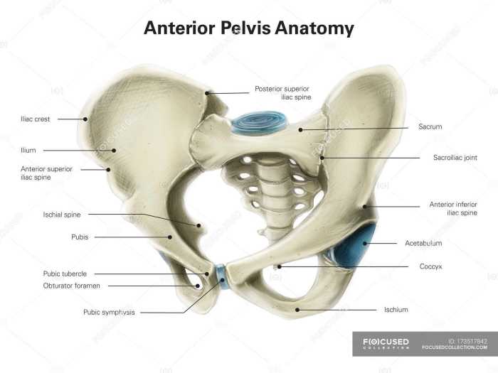
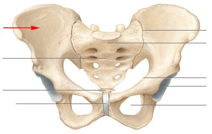
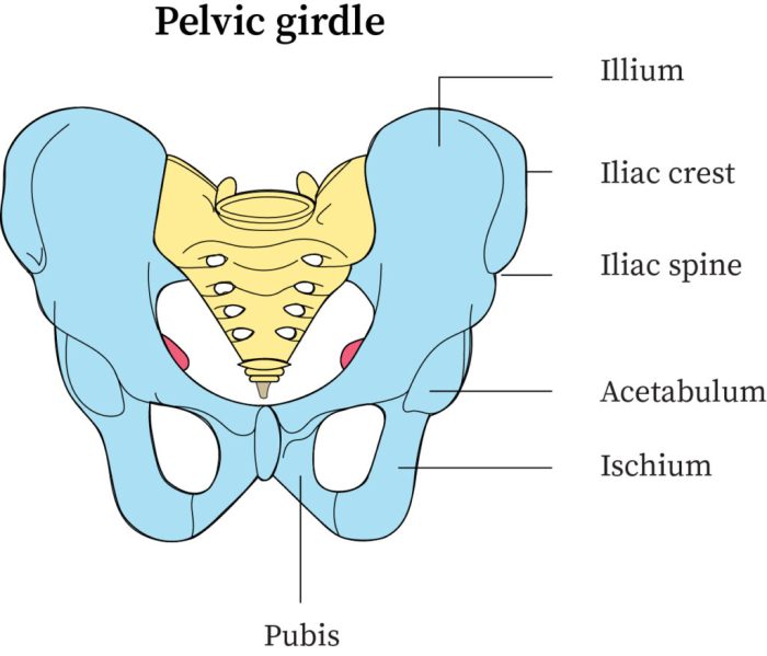
The pelvis, also known as the pelvic girdle, is a bony structure that forms the lower part of the trunk. It consists of two large, symmetrical bones called the hip bones, also known as the innominate bones.
The Hip Bones
- Shape and Size:The hip bones are large, flat, and fan-shaped bones that form the lateral and anterior walls of the pelvis.
- Location:They are located on either side of the sacrum and are connected to each other anteriorly by the pubic symphysis and posteriorly by the sacroiliac joints.
- Role:The hip bones provide stability and support to the pelvic girdle and transmit weight from the upper body to the lower limbs. They also serve as attachment points for muscles that control movement of the lower extremities.
Minor Bones of the Pelvis

In addition to the major pelvic bones, several smaller bones contribute to the structure and stability of the pelvis. These minor bones are located at the joints between the major bones and help to articulate and strengthen the pelvic ring.
Coccyx
- A small, triangular bone located at the inferior end of the sacrum.
- Composed of four fused vertebrae.
- Provides attachment for muscles and ligaments that support the pelvic floor.
Sacrococcygeal Joint
- Connects the coccyx to the sacrum.
- A synovial joint that allows for limited movement between the two bones.
Pubic Symphysis
- A cartilaginous joint between the pubic bones.
- Provides flexibility and shock absorption during childbirth.
- Contains the interpubic disc, which is a fibrocartilaginous structure that helps to maintain the stability of the joint.
Sacroiliac Joints, Bones of the pelvis quiz
- Joints between the sacrum and the ilium on each side.
- Strong, synovial joints that allow for limited movement.
- Transmit weight from the spine to the lower limbs.
Pelvic Joints

The pelvis is a ring-shaped structure composed of several bones that connect to form a stable and flexible framework. The joints of the pelvis play a crucial role in supporting the weight of the body, facilitating movement, and protecting the pelvic organs.
There are three major joints in the pelvis:
- Sacroiliac joint:This joint connects the sacrum to the ilium on each side. It is a synovial joint that allows for limited gliding and rotation movements.
- Pubic symphysis:This joint connects the two pubic bones anteriorly. It is a cartilaginous joint that permits some movement during childbirth and shock absorption.
- Hip joint:This joint connects the femur to the acetabulum of the pelvis. It is a ball-and-socket joint that allows for a wide range of motion, including flexion, extension, abduction, adduction, and rotation.
Ligaments and muscles play a vital role in stabilizing the pelvic joints. The strong ligaments that surround the joints provide support and limit excessive movement, while the muscles that cross the joints help to control movement and maintain stability.
Pelvic Muscles
The pelvis is a stable and mobile structure that supports the abdominal organs, provides attachment for the lower limbs, and facilitates childbirth. The stability and mobility of the pelvis are due in part to the muscles that attach to the pelvic bones.
Ace your bones of the pelvis quiz with ease! For a quick break, check out the ohio cdl air brake test to brush up on your knowledge. Afterward, return to the pelvis quiz and conquer those tricky questions like a pro!
Major Pelvic Muscles
- Iliac Muscle:
- Origin:Iliac fossa
- Insertion:Sacroiliac joint
- Function:Flexes the thigh at the hip joint
- Psoas Major Muscle:
- Origin:Lumbar vertebrae and intervertebral discs
- Insertion:Lesser trochanter of the femur
- Function:Flexes the thigh at the hip joint
- Obturator Internus Muscle:
- Origin:Obturator foramen
- Insertion:Greater trochanter of the femur
- Function:Laterally rotates the thigh at the hip joint
- Piiformis Muscle:
- Origin:Sacrum
- Insertion:Greater trochanter of the femur
- Function:Laterally rotates the thigh at the hip joint
- Coccygeus Muscle:
- Origin:Coccyx
- Insertion:Sacrum
- Function:Supports the pelvic floor
Clinical Significance
The pelvis, a ring-shaped structure, plays a crucial role in supporting the weight of the upper body, protecting internal organs, and facilitating childbirth. Injuries or disorders affecting the pelvic bones can have significant implications for an individual’s mobility, stability, and overall well-being.Common
pelvic injuries include fractures, dislocations, and ligament sprains or tears. These can result from high-impact events, such as falls or motor vehicle accidents, or from chronic conditions like osteoporosis. Pelvic fractures, in particular, can be severe and require immediate medical attention due to the risk of internal bleeding, nerve damage, and organ injury.Imaging
techniques, such as X-rays, computed tomography (CT) scans, and magnetic resonance imaging (MRI), play a vital role in diagnosing pelvic injuries. These tests help visualize the bones, soft tissues, and blood vessels to determine the extent and location of the injury.Surgical
intervention may be necessary to treat pelvic fractures, especially if they are unstable or involve the displacement of bone fragments. Surgical procedures aim to restore bone alignment, stabilize the pelvis, and minimize the risk of complications. Common surgical approaches include open reduction and internal fixation (ORIF), in which the bones are repositioned and held in place with plates, screws, or wires, and percutaneous fixation, which involves inserting pins or screws through the skin to stabilize the bones.
Questions and Answers
What is the largest bone in the pelvis?
The ilium
How many bones make up the pelvis?
Four (two hip bones and two sacral bones)
What is the function of the pelvic floor muscles?
Support the pelvic organs and maintain continence